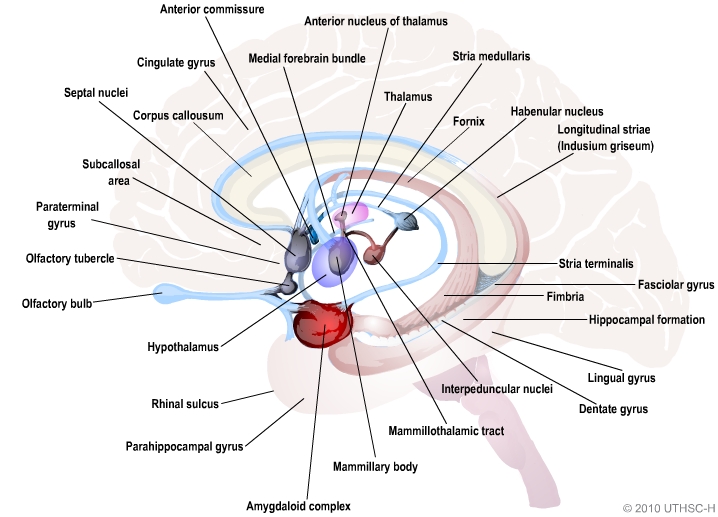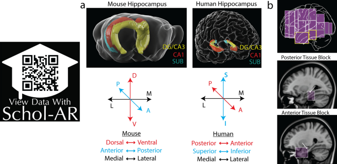

The hippocampus also receives direct monosynaptic projections from the cerebellar fastigial nucleus (Heath and Harper 1974).ĭiagram of hippocampal regions. In Macaca fascicularis, these inputs include the amygdala (specifically the anterior amygdaloid area, the basolateral nucleus, and the periamygdaloid cortex), the medial septum and the diagonal band of Broca, the claustrum, the substantia innominata and the basal nucleus of Meynert, the thalamus (including the anterior nuclear complex, the laterodorsal nucleus, the paraventricular and parataenial nuclei, the nucleus reuniens, and the nucleus centralis medialis), the lateral preoptic and lateral hypothalamic areas, the supramammillary and retromammillary regions, the ventral tegmental area, the tegmental reticular fields, the raphe nuclei (the nucleus centralis superior and the dorsal raphe nucleus), the nucleus reticularis tegementi pontis, the central gray, the dorsal tegmental nucleus, and the locus coeruleus. The hippocampus also receives a number of subcortical inputs. The subiculum is the final stage in the pathway, combining information from the CA1 projection and EC layer III to also send information along the output pathways of the hippocampus. In turn, CA1 projects to the subiculum as well as sending information along the aforementioned output paths of the hippocampus.

Region CA1 receives input from the CA3 subfield, EC layer III and the nucleus reuniens of the thalamus (which project only to the terminal apical dendritic tufts in the stratum lacunosum-moleculare). Region CA3 combines this input with signals from EC layer II and sends extensive connections within the region and also sends connections to region CA1 through a set of fibers called the Schaffer collaterals.

Perforant path input from EC layer II enters the dentate gyrus and is relayed to region CA3 (and to mossy cells, located in the hilus of the dentate gyrus, which then send information to distant portions of the dentate gyrus where the cycle is repeated). The lamellar concept is still sometimes considered to be a useful organizing principle, but more recent data, showing extensive longitudinal connections within the hippocampal system, have required it to be substantially modified.
#Hippocampus anatomy ca series#
This observation was the basis of his lamellar hypothesis, which proposed that the hippocampus can be thought of as a series of parallel strips, operating in a functionally independent way. The perforant path-to-dentate gyrus-to-CA3-to-CA1 was called the trisynaptic circuit by Per Andersen, who noted that thin slices could be cut out of the hippocampus perpendicular to its long axis, in a way that preserves all of these connections. Subicular neurons send their axons mainly to the EC. Pyramidal cells of CA1 send their axons to the subiculum and deep layers of the EC. Pyramidal cells of CA3 send their axons to CA1. Granule cells of the DG send their axons (called "mossy fibers") to CA3. There is also a distinct pathway from layer 3 of the EC directly to CA1. These axons arise from layer 2 of the EC, and terminate in the dentate gyrus and CA3. Most external input comes from the adjoining entorhinal cortex, via the axons of the so-called perforant path. The major pathways of signal flow through the hippocampus combine to form a loop.
#Hippocampus anatomy ca plus#
To refer to the hippocampus proper plus dentate gyrus and subiculum. Use the term "hippocampus proper" to refer to the four CA fields, and "hippocampal formation" Transition to the cortex proper (mostly the entorhinal area of the cortex). After this come a pair of ill-defined areas called the presubiculum and parasubiculum, then a After CA1 comes an area called the subiculum. The CA areas are all filled with densely packed pyramidal cells resembling those of the cortex.

NextĬome a series of Cornu Ammonis areas: first CA4 (which underlies the dentate gyrus), then CA3, thenĪ very small zone called CA2, then CA1. Proper, forming a pointed wedge in some cross-sections, a semicircle in others. Structure, a tightly packed layer of small granule cells wrapped around the end of the hippocampus The first of these, the dentate gyrus (DG), is actually a separate Starting at the dentate gyrus and working inward along the S-curve of the hippocampus means traversing a Basic circuit of the hippocampus, shown using a modified drawing by Ramon y Cajal.


 0 kommentar(er)
